


Introduction
Heredity appears to play a role in skin pigmentation, distribution of melanocytes, melanin content, and melanin activity. Variations in melanosome quantity, size, type, and arrangement within melanocytes are deemed responsible for the differences in skin color.
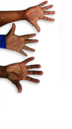
Racial (ethnic) differences in skin properties may explain racial disparities seen in dermatologic disorders and provide insight into appropriate differences in the management of these disorders. However, racial differences in skin have been minimally investigated by objective methods and the data are often contradictory. Objective methods studied include transepidermal water loss, water content, corneocyte variability, blood vessel reactivity, elastic recovery/extensibility, pH gradient, lipid content, surface microflora, microscopic evaluation of mast cell granules, and confocal microscopy.
Melanin / Melanosomes / Melanocytes
- Tyrosinase. In Caucasian melanocytes the melanosome-bound enzyme is largely inactive. Conversely melanosomes of Black melanocytes have up to 10 times more tyrosinase activity and produce up to 10 times more melanin than melanocytes derived from Caucasian skin.
However, the variation of levels of tyrosinase activity in melanocytes derived from black skin is not due to a greater abundance of tyrosinase gene activity but by a post-translational regulation of pre-existing enzyme.
Results from immunotitration experiments and Western immunoblots reveal that approximately equivalent levels of tyrosinase mRNA are present in white and black skin cell strains. In contrast, melanocytes derived from red-haired people contain low numbers of tyrosinase molecules and low levels of tyrosinase mRNA suggesting that transcriptional activity of the tyrosinase gene is suppressed. - PH. Data support that the rate of melanogenesis may be determined by differences in intracellular pH of melanocytes and melanosomes in Black and Caucasian skin types. The models suggest that melanosomes of Caucasian melanocytes are acidic, while those of Black individuals are closer to neutral pH. Since tyrosinase is inactive in an acidic environment, the enzyme is largely inactive in Caucasian melanosomes but optimal for melanin synthesis in Black melanosomes.
- Melanosome Distribution. Another distinguishing characteristic between light skin and dark skin is the pattern and distribution of melanosomes in the cytoplasm of keratinocytes.
A recent study using electron microscopy demonstrated that melanosomes within keratinocytes of African American skin have predominantly individually dispersed melanosomes (88.9%), Caucasian skin is predominantly distributed in membrane-bound clusters (84.5%) with clusters containing approximately 4 to 8 melanosomes and Asian skin has a combination of individual and clustered distribution of melanosomes with a proportion of 62.6% vs. 37.4%, respectively. - Rate of Degradation. As demonstrated in studies, the pattern of degradation of melanosomes may vary among ethnic groups. Transmission electron micrographs of skin taken from light and dark upper-arm skin biopsies demonstrate an enhanced degradation/reduction of melanosomes in light skin samples. In white skin melanosomes are confined to the lower epidermal layers, the stratum basale and stratum spinosum. Light skin lacks melanosomes in the stratum granulosum and stratum corneum, suggesting a differential processing of melanosomes between light skin and dark skin. Melanosomes in black skin are distributed throughout the epidermis with increased numbers in the basal layer. Dark skin showed retention of melanosomes throughout the epidermis, with melanosomes still apparent in the upper skin layers, including the stratum granulosum and stratum corneum.
- Melanosome Size. The present data indicate a size gradient of melanosomes in the keratinocytes and corneocytes across ethnicities with Africans having the largest melanosomes followed by Indians, Mexicans, Chinese, and Europeans
Electron microscopy analysis of mean melanosome size also revealed a progressive variation in size with ethnicity, with melanosomes in dark skin being the largest (1.44 +/- 0.67 microm(2) x 10-2) followed in turn by those in Asian skin (1.36 +/- 0.15 microm(2) x 10-2) and Caucasian skin (0.94 +/- 0.48 microm(2) x 10-2).
In addition, it was shown that the melanosomes that are individually distributed tend to have a larger size than the clustered melanosomes. - Shape. In light-skin type cells, increased melanin production resulted in a more elliptical shape of melanosomes. In melanosomes that constitutively produce more melanin, the tyrosine-induced melanogenesis caused enlargement in all dimensions.
- Melanogenesis. Research has also shown a greater rate of melanogenesis and higher melanin content in darker skin types with lighter skin types having approximately half as much epidermal melanin as the most darkly pigmented skin types as demonstrated in various studies both in vitro and in vivo.
Barrier Function
Although no large, multi-ethnic group studies have been performed looking at all of the skin barrier physiologic properties and their relation to clinical signs of disease, several small studies show that barrier function of the skin depends on the structure of the corneocytes, lipid content, and transepidermal water loss. In clinical practice, these small variations should play a role in ethnic-specific treatment regimens for common hyperpigmentation conditions.
- Stratum Corneum. In comparison with white skin, the black skin stratum corneum is equal in thickness and weight, but has more corneocyte layers and a more compact stratum corneum with greater intercellular cohesiveness: about twenty cell layers are observed in blacks versus sixteen layers in whites.
Although the size of the individual corneocytes is the same in black and white skin, the desquamation rate in certain locations is higher in black skin. This is likely because of increased desquamatory enzyme levels, such as cathepsin L2 in the lamellar granules of darker pigmented individuals, leading to an "ashy" manifestation.
A comparative study found that in Black subjects, a greater number of tape strippings were required for complete removal of the stratum corneum when compared to Whites with patients with skin types II/III requiring only 29.6 +/- 2.4 tape strippings to perturb the barrier, and skin type V/VI group requiring 66.7 +/- 6.9 tape strippings. Microscopically, Black skin demonstrated higher average numbers of stratum corneum layers than White skin.
Furthermore, while barrier function in skin type II/III recovered by approximately 20% by 6 hours and 55% by 48 hours, barrier function in skin type V/VI, independent of race, recovered more quickly, 43% and 72% at 6 and 48 hours, respectively.
Ethnic differences in stratum corneum pH have been explored and shown that pH regulates homeostasis in epidermal permeability. The most recent study examining pH of the cheek, forehead, and arm did not find any differences among African-Americans, Asian Indians, Caucasians, East Asians and Latinos. A prior study found a significantly lower pH in Black women compared to Caucasian women only after three tape strippings. Moreover, there were no significant differences in pH with further tape strippings suggesting pH is similar in both races within deeper layers of the skin. Another study by Warrier et al. demonstrated a lower baseline pH on the cheeks in Blacks but not on the legs. The overall results of these studies trend toward the hypothesis that Black skin has a lower pH compared to Whites with some variations according to body sites and depth of skin layers. - Transepidermal Water Loss. The majority of the evidence (six out of eight studies) indicates that transepidermal water loss is greater in Black skin compared with White skin. Transepidermal water loss measurements of Asian skin are inconclusive as they have been found to be equal to Black skin and greater than Caucasian skin, equal to Caucasian skin, and less than all other ethnic groups in different studies. Racial differences in water content, as measured by resistance, capacitance, conductance and impedance, are also inconclusive as the data are contradictory.
- Ceramide Levels. Black skin has the highest sebum content of all ethnicities followed by Whites, Hispanics and Asians. Black skin also has the lowest ceramide levels, which make black skin the most susceptible to transepidermal water loss and xerosis of the skin, when compared with other ethnic groups.
Photoprotective Properties / Response to UV Radiation
It is well documented in various studies that melanin acts as a neutral density filter that protects the skin from damage by absorbing and deflecting rays of UV light, and it is the source of skin pigmentation.
A study found that five times as much ultraviolet light (UVB and UVA) reaches the upper dermis of Caucasians as that of blacks. The main site of UV filtration in Caucasians is the stratum corneum, whereas in blacks it is the malpighian layer of the epidermis.
Another study showed that melanin content correlated inversely with cyclobutane pyrimidine dimers (CPD markers of DNA damage). This study demonstrated that the correlation coefficient was significantly higher in the lower epidermis compared to the upper epidermis in dark skin. In contrast, fair skin demonstrated similar CPD levels in both the upper and lower epidermis.
The melanin content, packaging and distribution of melanosomes can impact the degree of photoprotection across racial groups. Larger, individually dispersed stage-lV melanosomes (predominantly in Black skin) have a higher melanin content and absorb more UV light than aggregated, smaller melanosomes with less melanin content (predominantly in White skin).
These data are consistent with epidemiologic evidence and they also may indicate why blacks are less disposed to skin cancer, photoaging, phototoxic drug responses and are less susceptible to acute and chronic actinic damage.
>
>
>
>
SKIN TYPES
Evolution of Skin Colour
Racial Differences in Skin Properties
Fitzpatrick Scale
Fitzpatrick Skin Type Calculator