


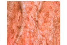
Age spots on a 63 year old woman's hand
Liver spots, also called lentigines or lentigos, age macular 1- to 3-cm, hyperpigmented, well-circumscribed lesions, are sharply defined, rounded, brown or black, flat patches of skin. They vary in color from light yellow to dark brown, and they often have a variegated appearance. They are usually seen on the arms, face and frequently on the back of the hands. One may appear by itself, or as a few clustered together. The term “liver spot” is actually not an appropriate name for this condition since these spots are not caused by liver problems or liver disease. In reality, these spots are not an indication of age but they do tend to develop over time and with repeated sunlight exposure. White or Asian persons are most likely to develop solar lentigines, especially those with skin types I to III. For the most part, people that do not tan, or sunburn, rather easily are at greatest risk to have this disorder. It is thought that the UV radiation in sunlight triggers mutations which, in turn, cause a local proliferation of basal melanocytes and a subsequent increase in the production of melanin. It is this increase in melanin that produces the so-called age spots by expanding the epidermis (top surface layer) with more pigment, developing what looks like a large freckle. In most cases, they are totally harmless.
The mechanisms underlying this type of hyperpigmentation process have recently been elucidated by Imokawa and coworkers. Besides a two-fold increase of TYR-positive melanocytes in lesional skin compared to perilesional skin, they demonstrated the existence of a molecular network in which increased expression of the ET-1/ET(B)R cascade and higher expression of SCF in lesional skin as well as cross-talk between these two signaling pathways following UV exposure play an important role in the mechanisms underlying LS.
Systemic disorders presenting with multiple lentigines may include Peutz-Jeghers syndrome (gastrointestinal hamartomas; buccal, lip, perioral, or digital macules; onset at birth or early childhood), LEOPARD syndrome (multiple lentigines, electrocardiogram abnormalities, ocular hypertelorism, pulmonic stenosis, abnormal genitalia, retarded growth, and sensorineural deafness), and LAMB syndrome (multiple lentigines, atrial and/or mucocutaneous myxomas, myxoid neurofibromas, ephelides, and blue nevi). Solar lentigines must be differentiated from premalignant lesions, such as pigmented actinic keratoses or lentigo maligna. Pigmented lesions with rapid growth or change, associated symptoms (e.g., pain, itching, easy or recurrent bleeding, poor healing), atypical lesions, or those with features suspicious for melanoma should be biopsied. Full thickness excisional biopsy or punch biopsy (for large lesions or those on the face or cosmetically sensitive area) is an acceptable method of biopsy in these instances. Solar lentigines can be distinguished clinically from flat seborrheic dermatoses or pigmented actinic keratoses by the absence of epidermal hyperkeratosis. Biopsy may facilitate diagnosis in uncertain cases.
>
>
>
>
>
>
>
>
>
>
>
>
>
>
>
>
>
>
>
>
>
>
>
>
>
>
INTRODUCTION
PIGMENTARY DISORDER TYPES
Disorders of
Hyperpigmentation
Disorders of Hypopigmentation
COMMON PIGMENTARY DISORDERS
Age Spots/Liver Spots/Lentigos
Acanthosis Nigricans
Albinism
Cafe-au-Lait Macules
Ephelides (Freckles)
Erythema Dyschromicum Perstans (Ashy Dermatosis)
Familial Racial Periorbital Hyperpigmentation
Idiopathic Guttate Hypomelanosis
Leopard Syndrome
Linea Nigra
Melanoma
Melasma
Nevus (birthmarks/moles)
Parkinsons Disease
Phytophotodermatits
Pityriasis Alba
Poikiloderma of Civatte
Postinflammatory
Hyperpigmentation & Hypopigmantation
Seborrheic Keratoses
Sturge-Weber Syndrome
Substance Induced
Hypermelanosis
Tinea Nigra/Tinea Versicolor/Pityriasis
Versicolor
Vitiligo
Waardenburg Syndrome

<< Previous: Disorders of Hypopigmentation