


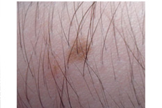
Nevus on arm
Birthmarks are areas of discolored and/or raised skin that are apparent at birth or within a few weeks of birth. Birthmarks are made up of malformed pigment cells or blood vessels. About 10 in every 100 babies have vascular birthmarks (birthmarks made up of blood vessels). Although the cause of birthmarks is not known, most of them are benign (non-cancerous) and do not require treatment.
The following are the most common types of vascular birthmarks:
Pigmented birthmarks
The discoloration of the skin will appear smooth and flat. These spots are known by several names, including Nevus of Ota, characterized by bluish discoloration of the facial skin and sometimes the white part of the eye (sclera); Mongolian spots, which are bruised or bluish in color, typically appearing on buttocks; café-au-lait spots, which are light brown; and typical moles, which are also called nevi, which may be flesh-colored to light to dark brown. Moles should be monitored for bleeding, color, shape or size changes, or itching.
Macular stains
These appear anywhere on the body, appearing as pink to red marks, but they flat and unelevated. Macular stains are the most common type of vascular (from blood vessels) birthmark. These marks can come in two forms: angel kisses, which may appear on the forehead and eyelids and typically disappear after age two; or stork bites, which will appear on the back of the neck and can last into adult years. Because these marks are often mild, there is no treatment necessary.
Hemangioma
Hemangiomas are growths composed of many tiny blood vessels bunched together and vary in severity. Typically, this birthmark is just a small mark on the skin of the face, trunk or extremities. However, in some children, hemangiomas can be large and grow rapidly through the first year of life, usually for about six to nine months. There are two types of hemangiomas: strawberry (or superficial) hemangiomas, which are slightly raised and can appear anywhere on the body; or cavernous (deep) hemangiomas, which are deeper birthmarks characterized by a bluish color. Fortunately, most hemangiomas will go away on their own. They gradually lose this red color and also shrink. 50% resolve by age five, 70% by age seven and 90% by age nine. Reasons to treat hemangioma include problems with functions (such as sight, eating, hearing or defecation), ulceration, bleeding or pain. If medically indicated, hemangiomas can be treated in different ways, each of which carries its own risks. Corticosteroid medication can be injected or taken orally. Risks associated with corticosteroid medication include high blood pressure, high blood sugar, poor growth, or cataracts. Certain hemangiomas can also be treated with lasers to stop them from growing and facilitate resolution. Rare risks associated with that treatment include ulceration and scarring. In some cases, a hemangioma can also be removed with surgery.
Port wine stains
Port wine stains are caused by abnormal development of blood vessels (capillaries) and last a lifetime. The port wine stain (also known as nevus flammeus) appears as a flat, pink, red, or purple mark, often occuring on the face, trunk, arms, or legs and continues to grow as the child grows. Port-wine stains do not go away and often require treatment if located on the eyelid or forehead. Port wine stains present on eyelids are thought to pose an increased risk of glaucoma. Physicians have tried many ways to treat port wine stains, including radiation, tattooing, freezing, dermabrasion, or sclerotherapy. Laser therapy is currently the treatment of choice, as it is the only method that destroys capillaries in the skin without causing damage to the rest of the skin. Port wine stains may be seen in certain medical disorders, including Sturge-Weber Syndrome, whose symptoms include port wine stains on the face, vision problems, convulsions, mental retardation, and perhaps even paralysis; and Klippel-Trenaunay Syndrome in which a limb has a trio of port wine stains, varicose veins, and/or too much bone and soft tissue growth. Each of these syndromes is very rare.
>
>
>
>
>
>
>
>
>
>
>
>
>
>
>
>
>
>
>
>
>
>
>
>
>
>
INTRODUCTION
PIGMENTARY DISORDER TYPES
Disorders of
Hyperpigmentation
Disorders of Hypopigmentation
COMMON PIGMENTARY DISORDERS
Age Spots/Liver Spots/Lentigos
Acanthosis Nigricans
Albinism
Cafe-au-Lait Macules
Ephelides (Freckles)
Erythema Dyschromicum Perstans (Ashy Dermatosis)
Familial Racial Periorbital Hyperpigmentation
Idiopathic Guttate Hypomelanosis
Leopard Syndrome
Linea Nigra
Melanoma
Melasma
Nevus (birthmarks/moles)
Parkinsons Disease
Phytophotodermatits
Pityriasis Alba
Poikiloderma of Civatte
Postinflammatory
Hyperpigmentation & Hypopigmantation
Seborrheic Keratoses
Sturge-Weber Syndrome
Substance Induced
Hypermelanosis
Tinea Nigra/Tinea Versicolor/Pityriasis
Versicolor
Vitiligo
Waardenburg Syndrome

<< Previous: Melasma
Next: Parkinsons Disease >>