


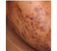
Postinflammatory hyperpigmentation from acne
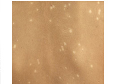
Postinflammatory hypopigmentation from psoriasis
Irritation from therapeutic interventions such as topical retinoids or benzoyl peroxide or other trauma to the skin like blisters and thermal burns may result in pigmentary changes.
Postinflammatory hyperpigmentation and hypopigmentation can also result from papulosquamous diseases such as allergic contact dermatitis and lichen planus or vesiculobullous diseases such as bullous pemphigoid and herpes zoster and common Inflammatiory cutaneous disorders like eczema, psoriasis and acne. Postinflammatory hyperpigmentation may also occur after laser therapy for other pigmented skin lesions, and may be transient or long lasting.
After cutaneous trauma or inflammation, melanocytes can react with normal, increased, or decreased production of melanin. Although the actual pathogenesis is unknown, it is thought that both hyperpigmentation and hypopigmentation result from cytokines and inflammatory mediators from keratinocytes, melanocytes, and inflammatory cells that are released in an inflammatory process in the skin. These include leukotriene (LT), prostaglandins (PG), and thromboxane (TXB). PGE1 and PGE2 increase melanogenesis strongly while PGA1 and PGD2 represent strong inhibitors Leukotrienes such as LTC4 and LTD4, are metabolites of the lipoxygenase pathway and are able to induce the proliferation of melanocytes in vitro. Histamine, another inflammatory agent, may activate melanogenesis via proteinase A activation.
In vitro studies have shown that LT-C4, in addition to LT-D4, PG-E2, and TXB-2, stimulate human melanocyte enlargement and dendrocyte proliferation. Also, LT-C4 significantly increases tyrosinase activity in cultured melanocytes and also increases mitogenic activity of melanocytes. In vitro studies have also shown that transforming growth factor-alpha and LT-C4 stimulate movement of melanocytes. These mediators and cytokines are thought to play an important role in the pathogenesis of postinflammatory hyperpigmentation.
The pathogenesis of postinflammatory hypopigmentation is believed to be secondary to melanocyte cell-surface expression of intercellular adhesion molecule (ICAM)-1 induced by inflammatory mediators such as interferon-gamma, tumor necrosis factor (TNF)-alpha, TNF-beta, interleukin (IL)-6 and IL-7. The theory is that this may lead to leukocyte-melanocyte attachments, with the final result being innocent bystander destruction of melanocytes.
The skin color of the hyperpigmentation is related to the location of the melanin. It appears brown when it is in the epidermis whereas blue to bluish-gray if it is in the dermis. With the use of a Wood’s lamp, the location of the increased melanin can be determined. The epidermal component is enhanced, whereas the dermal component becomes unapparent in hyperpigmentation. Hypopigmentation is accentuated with Wood’s lamp when it is due to melanocytopenic conditions such as vitiligo and piebaldism, in contrast to melanopenic conditions such as postinflammatory hypopigmentation. It should be noted, however, that the Wood’s lamp technique is sometimes not helpful in persons of color because of optical factors.
Treating the underlying cause of postinflammatory hyper- and hypopigmentation is an important aspect of preventing further dyschromia. Treatment modalities for postinflammatory hyperpigmentation include cosmetic cover-ups, hydroquinones, kojic acid, topical steroids, topical retinoids, chemical peels, and Q-switched ruby and Nd:YAG lasers. Postinflammatory hyperpigmentation tends to become darker and more noticeable with sun exposure so sun protection and broad-spectrum sunscreens are an essential part of management. For postinflammatory hypopigmentation, treatment modalities include cosmetic cover-ups, topical or oral Photo chemotherapy Ultraviolet A (PUVA) therapy, narrow-band UVB phototherapy, topical steroids, and skin grafts. Pigment loss is frequently not permanent, but it may take a long time to repigment.
>
>
>
>
>
>
>
>
>
>
>
>
>
>
>
>
>
>
>
>
>
>
>
>
>
>
INTRODUCTION
PIGMENTARY DISORDER TYPES
Disorders of
Hyperpigmentation
Disorders of Hypopigmentation
COMMON PIGMENTARY DISORDERS
Age Spots/Liver Spots/Lentigos
Acanthosis Nigricans
Albinism
Cafe-au-Lait Macules
Ephelides (Freckles)
Erythema Dyschromicum Perstans (Ashy Dermatosis)
Familial Racial Periorbital Hyperpigmentation
Idiopathic Guttate Hypomelanosis
Leopard Syndrome
Linea Nigra
Melanoma
Melasma
Nevus (birthmarks/moles)
Parkinsons Disease
Phytophotodermatits
Pityriasis Alba
Poikiloderma of Civatte
Postinflammatory
Hyperpigmentation & Hypopigmantation
Seborrheic Keratoses
Sturge-Weber Syndrome
Substance Induced
Hypermelanosis
Tinea Nigra/Tinea Versicolor/Pityriasis
Versicolor
Vitiligo
Waardenburg Syndrome

<< Previous: Poikiloderma of Civatte
Next: Seborrheic Keratoses >>