


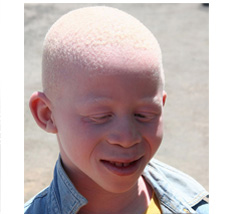
Child with albinism
Albinism refers to a set of distinct genetic disorders characterized by either a lack of pigment or pigment dilution. Albinism is a rare inherited autosomal recessive trait that is characterized by a total or partial lack of melanin in the skin. It is caused by a gene abnormality that leads to the absence of an enzyme that produces melanin. There are a number of characteristic phenotypic expressions as a result of hundreds of distinct gene mutations. The key feature of albinism is that melanocytes are normal in number and size but there is a defect in the synthesis of melanin. Immature melanosomes (stage I, II and III) are observed by electron microscopy. In Chédiak-Higashi syndrome, giant lysosomes are detected in the skin, leukocytes and other organs.
Albinism has many different forms, but most people who have this condition have pale skin, hair, and eyes. Melanin also creates eye color, and serves as a filter that prevents too much light from entering the eye. Since they lack melanin in their eyes, albinism affects not only eye pigmentation, but visual acuity, as well. People with albinism typically test poorly, within the 20/60 to 20/400 range. With little skin pigmentation, they also sunburn easily and are more prone to skin cancer. Additionally, two forms of albinism, with approximately 1 in 2700 most prevalent among people of Puerto Rican origin, are associated with mortality beyond melanoma-related deaths.
There are approximately ten different types of oculocutaneous albinism, which is mostly an autosomal recessive disorder. Certain ethnicities have higher incidences of different forms. For example, the most common type, called oculocutaneous albinism type 2 (OCA2), is especially frequent among people of black African descent. It is an autosomal recessive disorder characterized by a congenital reduction or absence of melanin pigment in the skin, hair and eyes. The estimated frequency of OCA2 among African-Americans is 1 in 10,000, which contrasts with a frequency of 1 in 36,000 in white Americans. In some African nations, the frequency of the disorder is even higher, ranging from 1 in 2,000 to 1 in 5,000. Another form of Albinism, the "yellow oculocutaneous albinism", appears to be more prevalent among the Amish, who are of primarily Swiss and German ancestry. People with this IB variant of the disorder commonly have white hair and skin at birth, but rapidly develop normal skin pigmentation in infancy.
Albinism occurs in two variants of hypo pigmentation.
1. Ocular albinism is a melanin by dysfunction that is limited to the eyes. This condition is an X-linked disorder.
2. Oculocutaneous albinism is a melanin synthesis defect that involves the eyes, skin, and hair, it predisposes to actinic keratosis, basal and squamous cell carcinoma, and malignant melanoma because of sensitivity of the skin to sunlight. Inheritance is almost often autosomal recessive.
Oculocutaneous albinism is often subclassified as:
a. Tyrosianse-negative albinism, which is failure of conversion of tyrosine to dihyrdropheylalanine (DOPA), an intermediary in melanin synthesis.
b. Tyrosinase-positive albinism. The mechanisms of deficient melanin synthesis are unknown.
Oculocutaneous albinism (OCA) can also be classified by the causative genes into OCA1, OCA2, OCA3 and OCA4. It is also seen as a symptom of hereditary diseases including Hermansky- Pudlak syndrome and Chédiak-Higashi syndrome.
1. OCA1
This is caused by tyrosinase gene mutation. OCA1 is classified into subtypes including OCA1A, in which tyrosinase activity is completely lost from such mutation (this type used to be classified as tyrosinase negative OCA), and OCA1B, in which some tyrosinase activity remains. All OCA1 subtypes are autosomal recessively inherited.
When melanin is not synthesized, as is the case in OCA1A, the skin appears white to pink, and the hair is white from birth.The skin is easily sunburned, and sun-exposed areas of the body are easily injured by UVR and are prone to malignant tumor (e.g., basal cell carcinoma, squamous cell carcinoma, malignant melanoma). The iris and choroid membrane are blue, and the ocular fundus is pink; the eyes appear blue when lit edgeon, and pink when lit head-on (pink-eye). Patients have a characteristic facial expression of squinting and looking out of the corner of the eyes, from photophobia and impaired eyesight that cannot be corrected. Horizontal nystagmus may be present.
When melanin is scant but present, pigment may gradually appear in hair and skin as the patient grows, although it is impossible to distinguish these cases from OCA1A at birth.
Oculocutaneous albinism (OCA) Type I is divided into two subtypes, A and B. Both are due to varying defects in the tyrosinase gene, leading to mild phenotypic variations in the disorder. OCA 1A is more severe than OCA 1B in that there is a complete lack of tyrosinase. This manifests as a congenital absence of pigment of the skin and eyes, which leads to impaired visual acuity as well as other neurological abnormalities. OCA IB differs in that markedly reduced tyrosinase activity is present, accounting for the visible amount of pigment that develops in the skin, hair, and eyes after birth. Thus, these patients are able to tan
.
2. OCA2
OCA2 is caused by mutation in the P protein gene on chromosome. It is autosomal recessive. In mice, the P protein works to convey tyrosine to melanosomes; however, the functions of human P protein have not been clarified. Pigment may be largely or completely absent at birth; it is impossible to distinguish OCA2 from OCA1 only by the clinical symptoms. In OCA2 the eye color is bluish gray and the hair is pale yellow to blonde; both come to contain more pigment as the patient ages.
OCA 2 is due to mutations in the P gene and can vary from a strikingly albino appearance to nearly normal pigmentation.
3. OCA3
OCA3 is caused by genetic mutation in TRP-1 (tyrosinaserelated protein 1), which controls melanin synthesis. It tends to occur in patients of African descent. The skin color is reddish brown, and the hair is light reddish brown to red. Eye symptoms do not usually occur.
OCA 3 is interesting in that it has been described only in ethnic populations, particularly blacks. Patients present with brown skin, brown hair, and blue or brown eyes. They commonly have decreased visual acuity and nystagmus as in other forms of oculocutaneous albinism. The defect has been localized to the tyrosinase related protein 1 (TRP-1) gene, whose function as yet remains poorly characterized. Studies suggest that a malfunction of this protein results in brown rather than black pigment formation, leading to its characteristic phenotype.
4. OCA4
OCA4 is caused by abnormality in the membrane-associated transporter protein (MATP). OCA4 is mainly seen in patients of African or Japanese ancestry. In Japan, it occurs with the second most frequency, after OCA1. Pigment is present in the skin in small amounts. The hair is light yellow in many cases; however, there are some cases in which the hair is brown The eyes are blue, gray or reddish brown. Nystagmus is found in about half of all cases.
5. Hermansky-Pudlak syndrome (HPS)
Some causative genes that are thought to be associated with intracellular protein transport have been identified in Hermansky- Pudlak syndrome (HPS). HPS is classified by the causative genes into four subtypes: HPS1, HPS2, HPS3 and HPS4. It is autosomal recessive. Pigment appears in the skin and hair to some extent. Mortality is increased in patients with Hermansky-Pudlak syndrome and. Pulmonary fibrosis or granulomatous colitis may occur as a complication by deposition of ceroid-lipofuscin. There is a hemorrhagic tendency in HPS, which manifests as susceptibility to bruising and nasal or gingival hemorrhaging. Patients with Hermansky-Pudlak syndrome have a bleeding diathesis secondary to platelet dysfunction and also experience inflammatory bowel disease, cardiomyopathy, and renal disease. Patients with Chediak-Higashi syndrome are susceptible to infection and also can develop lymphofollicular malignancy.
Hermansky-Pudlak syndrome is a form of oculocutaneous albinism seen almost exclusively in Puerto Ricans, but has also been reported in Japanese individuals. It is distinguished from the other types by its concomitant bleeding diathesis and ceroid storage in the aerodigestive tract.
6. Chédiak-Higashi syndrome (CHS)
Abnormality of the LYST gene on chromosome 1 (1q42) disturbs the normal function of microtubules. It is autosomal recessive. The main symptoms are partial albinism from melanocyte trafficking failure and photosensitive disorder. The hair is red and the skin color is cream, although sun-exposed areas such as the face sunburn to a dark red. Neutrophilic immune compromise often leads to bacterial infection. Histopathologically, giant lysosome granules (peroxidase-positive) are found in the peripheral leukocytes. During exacerbation, lymphatic and histiocytic infiltrate is found in the systemic organs, and acute symptoms of pancytopenia occur. Symptomatic therapies are performed for infection. Bone marrow transplantation may also be conducted. The prognosis is poor; most patients with CHS die young in the so-called “accelerated phase,” which is a lymphoproliferation into various organs resulting in hemophagocytosis, infection and bleeding.
Diagnosis/examinations:
The maturity of melanosomes in melanocytes should be observed by electron microscopy. In the severest OCA1 cases, there are only immature melanosomes that lack melanin deposition (stage I or II). In OCA in which some pigment production remains, stage III melanosomes are seen and there may also be a few stage IV melanosomes. Prenatal diagnosis may be conducted. for the severest OCA1A cases. Identification of the affected gene is necessary for determination of subtype.
>
>
>
>
>
>
>
>
>
>
>
>
>
>
>
>
>
>
>
>
>
>
>
>
>
>
INTRODUCTION
PIGMENTARY DISORDER TYPES
Disorders of
Hyperpigmentation
Disorders of Hypopigmentation
COMMON PIGMENTARY DISORDERS
Age Spots/Liver Spots/Lentigos
Acanthosis Nigricans
Albinism
Cafe-au-Lait Macules
Ephelides (Freckles)
Erythema Dyschromicum Perstans (Ashy Dermatosis)
Familial Racial Periorbital Hyperpigmentation
Idiopathic Guttate Hypomelanosis
Leopard Syndrome
Linea Nigra
Melanoma
Melasma
Nevus (birthmarks/moles)
Parkinsons Disease
Phytophotodermatits
Pityriasis Alba
Poikiloderma of Civatte
Postinflammatory
Hyperpigmentation & Hypopigmantation
Seborrheic Keratoses
Sturge-Weber Syndrome
Substance Induced
Hypermelanosis
Tinea Nigra/Tinea Versicolor/Pityriasis
Versicolor
Vitiligo
Waardenburg Syndrome

<< Previous: Acanthosis Nigricans