


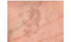
Tinea nigra on the palm of the hand
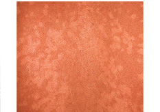
Pityriasis versicolor caused by fungus Malassezia furfur
Tinea versicolor is also called Pityriasis versicolor and is a superficial fungal infection of the stratum corneum. The causative agent for the pigmentary disorder is a lipophilic yeast that has been given several names. Currently, it is known as Pityrosporum ovale and is synonymous with Microsporon furfur, Malassazia furfur, and Pityrosporum orbiculare. It may induce enlarged melanosomes (pigment granules) within basal melanocytes resulting in hyperpigmented pityriasis versicolor. It is found throughout the world and in all races, although it favors tropical climates. It also tends to be more severe in the tropics.
It is characterised by scaly hypo- or hyperpigmented lesions on the body. The small circular lesions are most commonly found on the seborrheic areas of the trunk and may be very numerous. However, in tropical climates the infection can extend to the face, arms, and lower back. Affected areas do not tan well and become more noticeable in the summer or with tanning.
Tinea versicolor is not contagious. It has no racial or sexual preference. It seems to prefer hot, humid weather, which accounts for its prevalence in tropical climates or the summer season in temperate climates People who live in tropical areas may have tinea versicolor year-round. It is more common in adults, whose sebaceous glands are more active than those of children.
It has a distinctive clinical appearance. Classically, the disease presents as hypopigmented, dull white to tan scaly macules that are a few millimeters in diameter. Patches up to several centimeters in diameter are not unusual. Although clinically the acute eruption is unmistakable, a Wood’s lamp examination is sometimes necessary to distinguish this from vitiligo. Vitiliginous skin will appear stark white, whereas pityriasis versicolor-induced hypopigmented skin will have a slightly greenish hue. Microscopic examination of scales with potassium hydroxide reveals the classic appearance of hyphae and spores. The hypopigmentation resulting from the acute infection is the most distressing aspect of the disease in pigmented skins.
To date, a satisfactory explanation for the pathogenesis of the hypopigmentation remains elusive. UV light was thought to play a role but does not explain consistently the hypopigmentation seen in non-sun-exposed areas. In the past, the most compelling explanation for this disease was that the organism P. ovale has lipooxygenases that act on surface lipids, leading to the oxidization of oleic acid to azeleic acid. Azeleic acid was thought to block melanogenesis via competitive inhibition of tyrosinase. This was found to be erroneous as investigators discovered that the diacid had no depigmenting effect on normal skin. Although the enzymatic activity of the lipooxygenases is not in question, their effect currently is. Pityrosporum ovale’s activity on the unsaturated fatty acids found in sebum leads to the formation of lipoperoxidases. These enzymes are toxic only to abnormally active melanocytes. Their presence is thought to reduce DNA synthesis only in these melanocytes via mitochondrial damage, with subsequent depigmentation as an end result rather than direct prohibition of melanogenesis via hindrance of tyrosinase, as previously thought.
Eradication of the infection is difficult and recurrence rates within two years are 60-80%. Treatment of the acute disease consists of topical antifungal shampoos, creams, and lotions or systemic pulse antifungal therapy. After systemic or topical antifungal therapy, repigmentation is slow and recurrences are common. Chronic postinflammatory hypopigmentation can respond to topical or oral Psoralen UVA (PUVA) therapy or tar emulsion therapy, all of which may stimulate melanogenesis.
Tinea Nigra is a superficial fungal infection similar to Tinea/Pityriasis Versicolor and is caused by the fungus Hortaea Wemeckii. Sometimes other species of dematiaceous fungi, such as S araguata, may produce a similar clinical picture. It causes dark brown to black patches on the palms and soles of the feet that are usually painless. A pigmentary change in the skin results in a dark-colored macule due to the accumulation of a substance in the fungus that is similar to melanin.
Tinea Nigra usually occurs before adulthood and is most common to females. It appears to affect black population less than others.
>
>
>
>
>
>
>
>
>
>
>
>
>
>
>
>
>
>
>
>
>
>
>
>
>
>
INTRODUCTION
PIGMENTARY DISORDER TYPES
Disorders of
Hyperpigmentation
Disorders of Hypopigmentation
COMMON PIGMENTARY DISORDERS
Age Spots/Liver Spots/Lentigos
Acanthosis Nigricans
Albinism
Cafe-au-Lait Macules
Ephelides (Freckles)
Erythema Dyschromicum Perstans (Ashy Dermatosis)
Familial Racial Periorbital Hyperpigmentation
Idiopathic Guttate Hypomelanosis
Leopard Syndrome
Linea Nigra
Melanoma
Melasma
Nevus (birthmarks/moles)
Parkinsons Disease
Phytophotodermatits
Pityriasis Alba
Poikiloderma of Civatte
Postinflammatory
Hyperpigmentation & Hypopigmantation
Seborrheic Keratoses
Sturge-Weber Syndrome
Substance Induced
Hypermelanosis
Tinea Nigra/Tinea Versicolor/Pityriasis
Versicolor
Vitiligo
Waardenburg Syndrome

<< Previous: Substance Induced Hypermelanosis
Next: Vitiligo >>